We all want to monitor progression and many years ago it was suggested that TSPO is a marker of activated microglia and we have a large number of imaging studies claiming that using TSPO imaging we can see activated microglia in MS.
We however we published this paper.
Nutma E, Fancy N, Weinert M, Tsartsalis S, Marzin MC, Muirhead RCJ, Falk I, Breur M, de Bruin J, Hollaus D, Pieterman R, Anink J, Story D, Chandran S, Tang J, Trolese MC, Saito T, Saido TC, Wiltshire KH, Beltran-Lobo P, Phillips A, Antel J, Healy L, Dorion MF, Galloway DA, Benoit RY, Amossé Q, Ceyzériat K, Badina AM, Kövari E, Bendotti C, Aronica E, Radulescu CI, Wong JH, Barron AM, Smith AM, Barnes SJ, Hampton DW, van der Valk P, Jacobson S, Howell OW, Baker D, Kipp M, Kaddatz H, Tournier BB, Millet P, Matthews PM, Moore CS, Amor S, Owen DR. Translocator protein is a marker of activated microglia in rodent models but not human neurodegenerative diseases. Nat Commun. 2023 Aug 28;14(1):5247. doi: 10.1038/s41467-023-40937-z.
The title says it all so what do those the imaging community do?… Yep ignore the inconvenient information…
So rather than looking for something new and better, we plough on
So we get titles like this for example
Nasal xxxxxxx treatment of PIRA induces regulatory immunity, dampens microglial activation and stabilizes clinical progression in non-active secondary progressive MS. 2025 doi;2025.04.30.25326602.
So TSPO is picking up activated microglia in MS. Sadly by ignoring stuff it can mean SH1-In, SH1-Out, as it become hard to decipher what the imaging really means
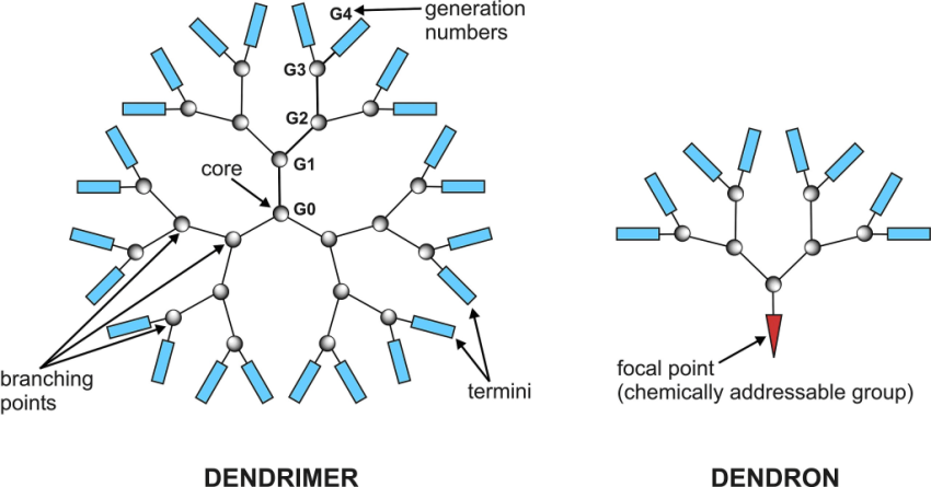
So here we have a paper with a dendrimer used for monitoring, Whats a dendrimer? Let’s find out.
Dendrimers are highly ordered, branched molecules that are ” symmetric about the core, and often adopt a spherical three-dimensional morphology.

By Olukin at English Wikipedia – Transferred from en.wikipedia to Commons., Public Domain, //commons.wikimedia.org/w/index.php?curid=3864829.
Here they use a fluorine-18-labeled (radioactive fluorine labeled for positron emission tomography…abit like an MRI) synthetic fourth-generation polyamidoamine hydroxyl dendrimer. Fourth-generation hydroxyl dendrimers have been demonstrated to penetrate regions of a dysfunctional, but not healthy, intact blood-brain barrier (BBB), where they are quickly and selectively taken up by activated myeloid cells
via fluid phase endocytosis, and rapidly cleared from tissue that does not contain these cells….(So alert bells ring here as surely they get taken up by macrophages and other cells too too. Indeed this is what was shown and also said that they detect “detect pathogenic microglia and peripheral CNS-infiltrating myeloid cells”). It can go up in EAE spinal cord and so is one of many different ways of showing blood brain barrier dysfunction. If you inhibit EAE with a drug the signal goes down because there are fewer lesions and less blood brain barrier disturbance.
Butter C, Baker D, O’Neill JK, Turk JL. Mononuclear cell trafficking and plasma protein extravasation into the CNS during chronic relapsing experimental allergic encephalomyelitis in Biozzi AB/H mice. J Neurol Sci. 1991;104:9-12.
Cellular traffic and plasma protein extravasation across the blood-brain and blood-spinal cord barrier (BBB) have been studied during chronic relapsing experimental allergic encephalomyelitis in Biozzi AB/H mice, using a simultaneous double radioisotope method. There was a general correlation between the clinical course of disease and BBB breakdown, including a resealing of the barrier during remission, although breakdown appeared slightly to precede clinical presentation. The brain was markedly less affected than the spinal cord and was only minimally involved in the relapse phase of disease.
So technologies evolve but is the take home message different?
I guess the question is does it offer advantage over things like paramagnetic detection of cells in lesions. The dendrimer was used to delivery inhibitors….interesting, but what will happen to the liver? The liver is full of myeloid cells that take up things. Bruton trysosine kinase inhibitors target myeloid cells and all of them seem to have a liver toxicity signal. Sadly with Nature type journals the legends aren’t suffieicntly informative to say what the pretty pictures are showing. Will it knock TSPO off its perch let’s see.
Kuo RC, Carlson ML, Reyes ST, Nagy SC, Kalita M, Alam IS, Malik N, Jackson IM, Acosta CJ, Falk IN, Azevedo EC, Zhang Y, Nichols L, Beinat C, Avci NG, Chattopadhyay M, Minami SS, Cleland JL, James ML. A radiolabeled dendrimer non-invasively identifies and tracks innate immune cell activation in a mouse model of experimental autoimmune encephalomyelitis. Nat Commun. 2026 Jan 30. doi: 10.1038/s41467-025-67907-x.
Multiple sclerosis (MS) is a chronic neurodegenerative disease driven by infiltration of activated innate immune cells into the central nervous system (CNS). Current imaging approaches for diagnosing and monitoring disease progression rely on structural lesions and cannot directly assess innate immune activity. Here, we describe a dendrimer positron emission tomography (PET) tracer, 18F-flurimedrimer (18F-FMD), for non-invasive, longitudinal tracking of activated myeloid cells. In an experimental autoimmune encephalomyelitis (EAE) murine model, 18F-FMD specifically detects myeloid activation at presymptomatic and symptomatic stages, with PET signal correlating with disease severity. Moreover, 18F-FMD sensitively captures therapeutic response to fingolimod (FTY720) and a CSF1R dendranib (H74DS3M8), both of which suppress immune cell activation and attenuate disease severity. These findings highlight the potential of 18F-FMD PET for specific, real-time monitoring of innate immune responses, and the applicability of the dendrimer in clinical settings for monitoring therapeutic efficacy, advancing the development of personalized, myeloid-targeted strategies for MS.
CoI None
Disclaimer: My views only..yep the post is about our
Source: multiple-sclerosis-research.org